;

Physiotherapy

Physiotherapists help people affected by injury, illness or disability through movement and exercise, manual therapy, education and advice. They maintain health for people of all ages, helping patients to manage pain and prevent disease. The profession helps to encourage development and facilitate recovery, enabling people to stay in work while helping them to remain independent for as long as possible.

Why Physiotherapy

Physiotherapy treatment approach prescribe not just medication and rest it also promote physical activity that's why physiotherapy is impotant for optimal health beacause it keeps you on the move.

Feel happy and Strong

Physiotherapy enables a person to regain his or her lost ablities due to any trauma or accident thus providing a mean to enjoy and live life more efficiently and Happilly.

Keep Moving Keep Active

Moving is life, everything which is stagnant does not last longer so if you want to add years in your life start exercising now.

Feel Healthy Look Healthy

Physical Activity enhance the performances of all body systems and enables you to do what ever you want by yourself with any aid or assisstance.

Thursday 28 September 2017

When to apply Ice or Heat for a Muscle Pain?

When to use ICE:

Ice should ALWAYS be used after an acute injury or trauma to any other area of the body.
Ice is a potent vasoconstrictor: cold causes the muscles of the body those lining the walls of our blood vessels, to constrict decreasing the swelling and pain associated with the body’s inflammatory response. As the vessels constrict, fewer inflammatory mediators seep into the area. In the case of musculoskeletal injuries, decreasing the inflammatory response decreases pain and prevents hyperimmune Une response in the area.
Ice should be applied at 15-20 minute intervals only, with at least an hour and a half in between icings.
Ice should be used for the first 48-72 hours following injury. Never use heat during this time.
Ice can also be used to alleviate pain associated with chronic back pain. Ice should be used after exercise, especially strenuous exercise, but NEVER before stretching or exercising. As ice causes increased muscle constriction and tension, its use before physical activity can lead to injury.


When to use HEAT:

Heat is used to relax and relieve tension associated with muscle stiffness and tension. Heat is best used to treat chronic, consistent back, neck and/or other musculoskeletal pain.
Heat can be applied before stretching and exercising to eliminate muscular stiffness and spasms. Warm towels or compresses work best.
These are just basic rules of thumb for treating back pain with heat or ice. If you have been diagnosed with an auto-immune disease, always discuss heat and ice therapies with your specialist. If you find that ice and/or heat seem to intensify your pain, avoid its use and consult with treating practitioner.


Friday 22 September 2017


Top 10 reasons to confirm Physiotherapists are the real Heroes.

1. Physiotherapist spends more time with the patients and listens to the problem carefully and tries to find the underlying cause by asking several questions from the patient.
2. Physiotherapist participates along with patient recovery with possible inputs throughout for a speedy recovery
3. The physiotherapist has to do physical treatment as well as gives the psychological boost to the patient.
4. A physiotherapist works with great team spirit, the physiotherapist works with coordination with the surgeon and physicians along with the whole medical team.
5. Physiotherapist respects other team members - Like a hero physiotherapist always respects other team members like a surgeon, physicians nurses, occupational therapists, speech therapists, prosthetic engineer and social workers of his team.
6. Physiotherapist ensures Slow, Steady and Sustainable Repair of the Body without harming the body.
7. Physiotherapist can maximize your movements
8. A physiotherapist helps to regain balance and coordination of mind with the body.
9. Physiotherapist offers safer ways to eliminate pain and improve functionality.
10. Physiotherapists avoid millions of invasive and expensive surgeries.
Doctors can give years to a life: but a Physiotherapist can give life to those years.
Share and spread the essence of Physiotherapy
Like and follow All about DPT - Doctor of Physiotherapy for more updates relating to physical therapy.


Friday 15 September 2017

For Athletes, the Risk of Too Much Water

Are we, with the best of intentions, putting young athletes at risk when we urge them to drink lots of fluids during steamy sports practices and games?
A new report about overhydration in sports suggests that under certain circumstances the answer is yes and that the consequences for young athletes can be — and in several tragic cases already have been — severe and even fatal.

Visit a practice for high school football, soccer or other team sports at this time of year, when temperatures can be high and early-season fitness marginal, and you are likely to see repeated water breaks and exhortations by the coaches and parents to drink up.

“A lot of people, including coaches, think that it is dangerous for athletes to get dehydrated, even a little dehydrated,” said Kevin Miller, an associate professor of athletic training at Central Michigan University in Mount Pleasant, Mich., and co-author of the new report.

The coaches and others worry that dehydration leads to muscle cramping and possibly heat illnesses, including serious heat stroke. So, hoping to keep their athletes healthy and safe, they press them to drink fluids before, during, and after a practice, whether the athletes feel thirsty or not.

And if an athlete should develop cramps or feel excessively hot during the workout, they are told to down even more fluids, and if the cramps continue, still more, “until, before you know it, a player will have drunk a gallon or two of fluid or even more,” Dr. Miller said, “which is something that we know actually happens.”

The problem with this situation is that, according to the latest science, dehydration during sports is rarely if ever dangerous, but overhydration undeniably is.

Last year, for instance, in a heartbreaking incident, a high school football player in Georgia experienced cramps during practice, and hoping to alleviate them, began gulping large amounts of water and Gatorade. By the end of the practice, he had swallowed about four gallons of fluid, according to media reports. Not long afterward, he collapsed at home and was rushed by helicopter to the hospital, where, several days later, he died.

At least two other high school football players are known to have died since 2008 from drinking too much fluid during and after a practice, Dr. Miller said. These players had developed a rare condition, he said, known formally as exercise-associated hyponatremia and less technically as water intoxication.

Hyponatremia occurs when someone consumes so much fluid that his or her body can’t rid itself of the surplus through sweating or urination. As a result, water levels rise in the bloodstream and sodium levels, diluted, fall. Osmosis then draws water from the blood into the surrounding cells of the body to equalize sodium levels there, and those cells begin to swell like water balloons. If this process occurs in the brain, it can be lethal.

Until recently, hyponatremia had been associated almost exclusively with marathon races and other prolonged endurance events, especially among slow racers, who tended to sweat little but drink copiously, often for hours on end. But as the new report, which presents updated hydration guidelines developed by a consortium of scientific experts, points out, exercise-associated hyponatremia “is now being reported in a more diverse set of sporting activities,” including half-marathons, sprint triathlons, Grand Canyon hikes, Bikram yoga classes, and, of course, team sport practices and games, especially football, at the professional, collegiate, and now high school level.

“What is sad is that every case” of exercise-associated hyponatremia “is preventable,” Dr. Miller said.

The key, he said, is for athletes to drink when they feel thirsty — not before and not after they feel sated. “You do not need to ‘stay ahead of your thirst,’ as many people think,” he said.

Listening to your “innate thirst mechanism” provides a safe and reliable guide to hydration, the new report concludes.

This strategy also should not increase players’ risks for cramping or heat illness, Dr. Miller said, since, “based on current evidence, it does not appear that dehydration directly contributes” to those problems.

During recent telling experiments that he directed, for instance, volunteers who exercised and sweated in the heat until they had become severely dehydrated were no more prone to muscle cramps than they had been at the start.

Similarly, if perhaps more surprising, other studies have found that being dehydrated does not increase athletes’ susceptibility to heart problems and that athletes who collapse from heat illness often are quite well-hydrated.

Instead, both cramping and heat problems seem to result from athletes pushing themselves too hard. Muscles cramp, Dr. Miller said, when a muscle is fatigued and begins to spasm, not when an athlete is dehydrated, while heat illnesses generally occur in athletes who are not physiologically acclimated to hot weather (a process that requires slowly increasing the length and intensity of workouts in the heat) and who continue to exercise even as they start to feel awful.

So, he said, “the best advice” about how to keep young athletes healthy during warm-weather practices and games, “is common sense.” Don’t urge athletes to drink if they aren’t thirsty. And don’t make them keep playing if they aren’t feeling well, he said.

If they complain of feeling too hot, have them sit in the shade and remove clothing. Take their temperature if they remain lethargic, and seek medical attention if it is much above normal. Immerse them in an ice bath, too, to rapidly lower body temperature. (Dr. Miller and his colleagues recently completed a study in which they found that football players who overheated could be submerged wearing full pads and uniforms and cool off almost as quickly as players dressed only in T-shirts and underwear, which could save precious minutes when a player seriously overheats.)

Above all, remind them, and, if needed, yourself, that the point of this enterprise is to have fun.


Why every athlete should do YOGA


As a private trainer and yoga instructor, I meet a lot of athletes and workout fanatics. When asked if yoga is part of their workouts, many athletes will tell me they don't have the time to add yoga to their already intense training schedules. Some say they don't see how "stretching and breathing" would be of any benefit to them. Others say they've discovered yoga while recuperating from an injury.

Why not enhance your performance and prevent injury by adding yoga to your training plan now? A well-rounded yoga practice includes dynamic flexibility training, core stabilization, strengthening and balance work. By focusing on these vital elements, yoga can help you recover faster after workouts, open up the tight areas that hinder performance, improve range of motion, and developmental focus and concentration.

"I've definitely noticed benefits in my triathlon training from yoga," said Heidi Resiert, a triathlete from San Diego. "My recovery time is much quicker, my shoulders feel stronger in the pool, and my muscles don't feel as tight after long runs or bike rides. I'm glad I found yoga and added it to my weekly workout routine. Not only do I feel stronger, but I also feel more confident that I will continue to be injury free."


Prevent Injury


Many sports, such as cycling and running, have very repetitive movements usually in one direction and in one plane of motion. These sports can develop certain muscle groups while ignoring others. Over time, this process causes imbalances in the muscles and joints leading to overuse injuries. For instance, tight hamstrings and hip flexors will cause the body to recruit from other joints, joints not intended for bearing extra loads.

Common overuse injuries among athletes include those involving the iliotibial band (ITB), knee, hamstrings, hip flexors, and shoulders. Often, these injuries are directly linked to lack of flexibility, poor core strength and misalignment. Yoga helps alleviate this tightness, builds a stronger center, and aligns the spine. In order to minimize and/or prevent injury, athletes should concentrate their efforts on these areas used most in endurance sports.

Even if athletes stretch pre- or post-workout, they are usually just stretching the muscles in the same direction and plane of motion in which they will be exercising. Yoga goes beyond simple stretching by working the muscles and joints through all ranges of motion--activating the little-used muscles that support the primary movers. The body must be worked through all three planes of motion in order to remain balanced and healthy. Yoga works not just in the sagittal plane but, in the frontal and transverse planes as well, ensuring well-rounded development.

Many yoga poses, such as Revolved Crescent, feature twisting motions in the transverse plane, essential to opening up tight obliques and lower backs. Balancing postures like Tree or Eagle are some of the most effective ways to correct muscle imbalances and poor body mechanics.


Another essential element in yoga is breath work or pranayama. The attention to breath during yoga can be considered one of the most important benefits to athletes. Learning to stay focused and centered through uncomfortable poses by concentrating on even inhalations and exhalations set up the athlete to stay focused during a race or challenging workout. The mind-body connection in yoga is essential to helping athletes develop mental acuity and concentration. In addition, yoga helps you to relax not just tight muscles, but also anxious and overstressed minds. Being more relaxed will also aid in athletic performance.


Where to Start

Yoga has been practiced for around 5,000 years and several schools of yoga have emerged over time. It can be overwhelming at first to find a style of yoga that resonates with you. If you are a competitive athlete, it is best to tailor your yoga practice to your training schedule. On a day where you are completing a long run, for example, you'll want relaxing, mellow yoga. If you have an off-day, a vigorous, dynamic class will help you build strength and endurance.

You can choose from dynamic styles like Ashtanga yoga and Power yoga that consist of a rigorous flowing series of poses synchronized with breath to produce internal heat and purifying sweat. Alternatively, Bikram yoga is a set series of 26 static poses performed in the 105-degree room. Iyengar focuses primarily on anatomical precision and alignment in poses, with an emphasis on healing the body and mind using postures. Anusara is a tantric-based system that combines alignment with awareness of energy flow in the body. Finally, there are restorative styles such as gentle Hatha and Yin yoga which feature longer holds.

Yoga helps the muscles, tendons, and ligaments move through a full range of motion, thus cultivating balance and core strength which is a huge benefit to athletes in their chosen sports. If you attend a few classes per week and/or a few 10-15 minute sessions at home, you will obtain fast results. A simple way to add in yoga is to perform your short sessions pre- or post-workout. Try it and see for yourself.


Pacemaker: The Basic Info


Faulty electrical signaling in the heart causes arrhythmias. A pacemaker uses low-energy electrical pulses to overcome this faulty electrical signaling. Pacemakers can:

  • Speed up a slow heart rhythm.

  • Help control an abnormal or fast heart rhythm.

  • Make sure the ventricles contract normally if the atria are quivering instead of beating with a normal rhythm (a condition called atrial fibrillation).

  • Coordinate the electrical signaling between the upper and lower chambers of the heart.

  • Coordinate the electrical signaling between the ventricles.

  • Pacemakers that do this are called cardiac resynchronization therapy (CRT) devices. CRT devices are used to treat heart failure.

  • Prevent dangerous arrhythmias caused by a disorder called long QT syndrome.

Pacemakers also can monitor and record your heart's electrical activity and heart rhythm. Newer pacemakers can monitor your blood temperature, breathing rate, and other factors and adjust your heart rate to changes in your activity.

Pacemakers can be temporary or permanent. Temporary pacemakers are used to treat temporary heartbeat problems, such as a slow heartbeat that's caused by a heart attack, heart surgery, or an overdose of medicine.

Temporary pacemakers also are used during emergencies. They're used until a permanent pacemaker can be implanted or until the temporary condition goes away. If you have a temporary pacemaker, you'll stay in a hospital as long as the device is in place.

Permanent pacemakers are used to control long-term heart rhythm problems. This article mainly discusses permanent pacemakers, unless stated otherwise.

Doctors also treat arrhythmias with another device called an implantable cardioverter defibrillator (ICD). An ICD is similar to a pacemaker. However, besides using low-energy electrical pulses, an ICD also can use high-energy electrical pulses to treat certain dangerous arrhythmias.


What Is a Pacemaker?

A pacemaker is a small device that's placed in the chest or abdomen to help control abnormal heart rhythms. This device uses electrical pulses to prompt the heart to beat at a normal rate.

Pacemakers are used to treat arrhythmias (ah-RITH-me-ahs). Arrhythmias are problems with the rate or rhythm of the heartbeat. During an arrhythmia, the heart can beat too fast, too slow, or with an irregular rhythm.

A heartbeat that's too fast is called tachycardia (TAK-ih-KAR-de-ah). A heartbeat that's too slow is called bradycardia (bray-de-KAR-de-ah).

During an arrhythmia, the heart may not be able to pump enough blood to the body. This may cause symptoms such as fatigue (tiredness), shortness of breath, or fainting. Severe arrhythmias can damage the body's vital organs and may even cause loss of consciousness or death.

A pacemaker can relieve some arrhythmia symptoms, such as fatigue and fainting. A pacemaker also can help a person who has abnormal heart rhythms resume a more active lifestyle.


For more information: http://bit.ly/GVf9gu

Acute Compartment Syndrome


Acute compartment syndrome of the leg is a surgical emergency which can lead to significant disability and even death if not diagnosed and treated early.

It is far more common in men than women, with a reported annual incidence of 7.3 per 100,000 and 0.7 per 100,000 respectively.

To understand the pathogenesis of compartment syndrome in the leg, one needs to appreciate that the deep fascia and long bones divide the leg into four distinct compartments - anterior, lateral, superficial posterior and deep posterior.

Under normal circumstances, the pressure inside each compartment is quite low (typically between 0 to 8 mmHg). This is well below the capillary closing pressure.

Unfortunately, the deep fascia is relatively inelastic, i.e. the volume of each compartment is fixed. Thus, even minor derangements in tissue homeostasis may result in rapid increases in compartmental pressure.

In this regard, elevation of tissue pressures may occur either because of increased compartmental contents (i.e. bleeding following trauma, edema secondary to nephrotic syndrome, or tissue destruction following snakebite); because of diminished compartment volume (i.e. after burns, or due to tight plaster casts); and very rarely, because of injuries to the microvasculature (i.e. in diabetes).

In practice, the commonest cause is a fracture of the tibial diaphysis, followed by blunt soft-tissue injury.

If the compartmental pressure rises above the capillary closing pressure, circulation shuts down, resulting in intra-compartmental ischemia and progressive necrosis of the muscles.

In turn, the skeletal muscles respond to the ischemia by releasing histamine-like substances which increase vascular permeability. Plasma leaks out of the capillaries, aggravating the tissue edema and worsening the ischemia.

Compartment syndrome of the leg is a clinical diagnosis, with the classical features being:

- Severe pain, typically increasing over time and often resistant to analgesics. In patients with a history of trauma, the pain is usually out of proportion to the apparent injury.

- Worsening of the pain when passively stretching the muscles within the affected compartment.

- A palpably tense compartment.

- Weakness and paresthesia of the areas supplied by nerves traversing the compartment. Note that these are late features and indicate the severe compromise.

It is uncommon for the compartment pressure to rise above the arterial pressure - thus pedal pulses and distal capillary filling are often normal. Pulselessness and diminished capillary filling are more likely due to arterial injury, or compression by a hematoma.

Note that non-classical presentations are common. In particular, pain is a highly unreliable symptom as it is subjective, difficult to elicit in patients with altered mentation, and sometimes absence in established compartment syndrome.

Where the clinical picture is equivocal, compartment pressure measurement may aid the decision-making process. Most authorities agree that an absolute resting pressure of over 30 to 45 mmHg, or within 30 mmHg of the diastolic blood pressure is suggestive of compartment syndrome.

Once the diagnosis is made, the most important step of the management is prompt decompression of the compartment via a fasciotomy.

All four compartments are usually decompressed, in order to avoid missing an affected compartment. This may be performed via a single incision or a double incision, which should extend across the entire length of the muscle and involve all layers of the skin to the deep fascia (as these may otherwise restrict expansion).

Following surgery, the wounds should be left open to allow the compartmental swelling to subside. In certain patients, repeated debridement to remove necrotic soft tissue may be necessary.

Wound closure may be achieved via several techniques. Split-thickness skin grafting is often employed. Delayed primary closure is an alternative, but may require an extended delay (to allow approximation with minimal or no skin tension). Vacuum-assisted wound closure is another option gaining popularity.

In the long term, these patients should receive physical therapy to regain function and facilitate rehabilitation.

During the acute stage, close observation for rhabdomyolysis, acute renal failure and sepsis is important.

The morbidity of acute compartment syndrome is directly related to the timing of decompression. When performed within 12 hours of onset, normal limb function is regained in 68% of patients. If delayed beyond this point, this declines to 8%.

Key long-term complications include residual numbness, peroneal nerve palsy, and limited motor function of the involved muscles.

Complications of fasciotomy include wound infections, decreased sensibility within the wound area, tethered tendons, and recurrent ulcerations within the wound closure area.


The main causes of mortality are sepsis and multiorgan failure.


Watch this video for more illustration.

Tuesday 29 August 2017

Cold Water Immersion for Athletes


Many athletes use `cold water immersion` post-exercise with the belief that it will assist in muscle recovery and improve performance... But does it?
Bret Contreras, a Strength & Conditioning Specialist (2015) evaluates the current evidence with regards to cold water immersion post-exercise and reviews its physiological effects below.


The key findings of this review were:
--------------------------------------------------
1) Cold water immersion substantially attenuated long-term gains in muscle mass and strength (Roberts et al, 2015)
2) Delayed and/or suppressed the activity of satellite cells and kinases in the mTOR pathway during recovery from strength exercise (Roberts et al, 2015)
3) Cold water immersion doesn’t appear to outperform a Placebo (Broatch et al, 2014)
4) It doesn’t seem improve sleep architecture (Robey et al, 2013)
5) A major review paper didn’t approve of it for treating muscle soreness (Bleakley et al, 2012)
6) A review paper concluded that cold water immersion benefited endurance athletes in terms of recovery, but not strength/power athletes (Chow et al, 2015)


Conclusions:
------------------
"The authors from the Roberts et al. (2015) study propose that regular deficits in acute hypertrophy signalling in muscle after cold water immersion accumulated over time, which in turn resulted in smaller improvements in strength and hypertrophy. The present findings contribute to an emerging theme that cold water immersion and other strategies (e.g., antioxidant supplements, non-steroidal anti-inflammatory drugs) that are intended to mitigate and improve resilience to physiological stress associated with exercise may actually be counterproductive to muscle adaptation.


This investigation offers the strongest evidence to date that using cold water immersion on a regular basis may interfere with training adaptations. No previous study has investigated the effect of cold water immersion on muscle hypertrophy after strength training.”


Its good news for endurance athletes, as cold water immersion seemed to have benefited endurance athletes in terms of recovery, but not strength/power athletes (Chow et al, 2015).


The other key findings from the review can be seen listed above!


Sunday 27 August 2017

What is Physiotherapy/Physical Therapy


Physical therapy (PT), mostly known as Physiotherapy, is a primary care specialty in western medicine that, by using mechanical force and movements [Bio-mechanics or Kinesiology], Manual therapy, exercise therapy, electrotherapy and various physical therapies who practice evidence based treatments, remediates impairments and promotes mobility, function, and quality of life through examination, diagnosis, prognosis, and physical intervention. It is performed by physical therapists (known as physiotherapists in many countries).

In addition to clinical practice, other activities encompassed in the physical therapy profession include research, education, consultation, and administration. Physical therapy services may be provided as primary care treatment or alongside, or in conjunction with, other medical services.


What Exactly Physiotherapists do?

Physical therapists (PTs) are highly-educated, licensed health care professionals who can help patients reduce pain and improve or restore mobility - in many cases without expensive surgery and often reducing the need for long-term use of prescription medications and their side effects.

Physical therapists can teach patients how to prevent or manage their condition so that they will achieve long-term health benefits. PTs examine each individual and develop a plan, using treatment techniques to promote the ability to move, reduce pain, restore function, and prevent disability. In addition, PTs work with individuals to prevent the loss of mobility before it occurs by developing fitness- and wellness-oriented programs for healthier and more active lifestyles.

Physical therapists provide care for people in a variety of settings, including hospitals, private practices, outpatient clinics, home health agencies, schools, sports and fitness facilities, work settings, and nursing homes. State licensure is required in each state in which a physical therapist practices.


According to the Olean Physical Therapy Profession there are five types of physiotherapy:


The field of physical therapy has sub-specialties in five distinct practice areas:
Orthopedic

Orthopedic physical therapy focuses on restoring function to the musculoskeletal system, including joints, tendons, ligaments and bones. Many sports injuries fall into this category. Treatment methods include stretching, strength training, endurance exercises, hot and cold packs, ultrasound, electrical muscle stimulation and joint mobilization.
Geriatric

Geriatric physical therapy focuses on the unique movement needs of older adults. This includes treatment for conditions such as arthritis, cancer, osteoporosis, Alzheimer's disease, joint replacement and balance disorders. The goal of geriatric physical therapy is to help restore mobility, reduce pain, accommodate physical limitations and increase physical fitness.
Neurological

Neurological physical therapy focuses on neurological conditions and impairments, such as Alzheimer's disease, brain injury, cerebral palsy, multiple sclerosis, Parkinson's disease, spinal cord injury and stroke. Treatment plans attempt to achieve the highest level of autonomous function for living as independently as possible for as long as possible. Physical therapists concentrate on teaching clients to adapt to visual, balance, mobility and muscle loss impairments for activities of daily living.
Cardiopulmonary

Cardiovascular and pulmonary physical therapy focuses on helping individuals who suffer from cardiovascular and pulmonary conditions, such as heart attacks, chronic obstructive pulmonary disease (COPD) and pulmonary fibrosis. The goal of this sub-specialty is to increase endurance and improve functional independence.
Pediatric

Pediatric physical therapy focuses on the unique needs of infants, toddlers, children and adolescents. Early detection is desirable when a child experiences problems that inhibit natural movement and learning. Physical therapy is used for children suffering from diseases or injuries, including:
acute injury
birth defects
developmental delays
genetic disorders (e.g., cerebral palsy)
head trauma
limb deficiencies
muscle diseases
orthopedic disabilities

Physical therapists use therapeutic exercise, modalities and treatments to help children alleviate pain, regain strength and range of motion, master proper body mechanics for balance and flexibility and improve gross and fine motor skills.

Two emerging sub-specialty practices in physical therapy are:

Vestibular Rehabilitation, which focused on balance problems caused by deficits in the inner ear, such as vertigo or dizziness.

Women's Health, which focused on relieving pelvic pain, reducing lymphatic swelling and controlling urinary incontinence.


10 Reasons why Physiotherapy if Beneficial

Physical therapy helps people of all ages who have medical conditions, illnesses or injuries that limit their regular ability to move and function.

A customized physical therapy program can help individuals return to their prior level of functioning, and encourage activities and lifestyle changes that can help prevent further injury and improve overall health and well being. Primary care doctors often refer patients to physical therapy at the first sign of a problem, since it is considered a conservative approach to managing problems. Wondering what makes physical therapy so important? In honor of Physical Therapy month in October, here are 10 ways it may benefit you:
Reduce or eliminate pain. Therapeutic exercises and manual therapy techniques such as joint and soft tissue mobilization or treatments such as ultrasound, taping or electrical stimulation can help relieve pain and restore muscle and joint function to reduce pain. Such therapies can also prevent pain from returning.

Avoid surgery. If physical therapy helps you eliminate pain or heal from an injury, surgery may not be needed. And even if surgery is required, you may benefit from pre-surgery physical therapy. If you are going into a surgery stronger and in better shape, you will recover faster afterwards in many cases. Also, by avoiding surgery, health care costs are reduced.

Improve mobility. If you’re having trouble standing, walking or moving—no matter your age—physical therapy can help. Stretching and strengthening exercises help restore your ability to move. Physical therapists can properly fit individuals with a cane, crutches or any other assistive device, or assess for orthotic prescription. By customizing an individual care plan, whatever activity that is important to an individual’s life can be practiced and adapted to ensure maximal performance and safety.

Recover from a stroke. It’s common to lose some degree of function and movement after stroke. Physical therapy helps strengthen weakened parts of the body and improve gait and balance. Physical therapists can also improve stroke patients’ ability to transfer and move around in bed so that they can be more independent around the home, and reduce their burden of care for toileting, bathing, dressing and other activities of daily living.

Recover from or prevent a sports injury. Physical therapists understand how different sports can increase your risk for specific types of injuries (such as stress fractures for distance runners). They can design appropriate recovery or prevention exercise programs for you to ensure a safe return to your sport.

Improve your balance and prevent falls . When you begin physical therapy, you will get screened for fall risk. If you’re at high risk for falls, therapists will provide exercises that safely and carefully challenge your balance as a way to mimic real-life situations. Therapists also help you with exercises to improve coordination and assistive devices to help with safer walking. When the balance problem is caused by a problem in one’s vestibular system, Physical therapists can perform specific maneuvers that can quickly restore proper vestibular functioning, and reduce and eliminate symptoms of dizziness or vertigo.

Manage diabetes and vascular conditions. As part of an overall diabetes management plan, exercise can help effectively control blood sugar. Additionally, people with diabetes may have problems with sensation in their feet and legs. Physical therapists can help provide and educate these patients on proper foot care to prevent further problems down the road.

Manage age-related issues . As individuals age, they may develop arthritis or osteoporosis or need a joint replacement. Physical therapists are experts in helping patients recover from joint replacement, and manage arthritic or osteoporotic conditions conservatively.

Manage heart and lung disease. While patients may complete cardiac rehabilitation after a heart attack or procedure, you also may receive physical therapy if your daily functioning is affected. For pulmonary problems, physical therapy can improve quality of life through strengthening, conditioning and breathing exercises, and help patients clear fluid in the lungs.

Manage Women’s Health and other conditions. Women have specific health concerns, such as with pregnancy and post-partum care. Physical therapists can offer specialized management of issues related to women’s health. Additionally, PT can provide specialized treatment for: Bowel incontinence, breast cancer, constipation, fibromyalgia, lymphedema, male pelvic health, pelvic pain, and urinary incontinence.

If you have questions about how physical therapy may help you or someone you care about, please don’t hesitate to ask your health care provider or local PT for information.


Friday 25 August 2017

Do you ever heard about OA?


The most common form of arthritis, affecting millions of people worldwide.Rheumatoid arthritis - the second most common form of arthritis - affects other parts of the body besides the joints.A joint disease that mostly affects cartilage,the slippery tissue that covers the ends of bones in a joint that breaks down and wears away over time.In a normal joints,a healthy cartilage is a firm,slippery tissue provides a smooth, gliding surface for joint motion and acts as a cushion. In OA, the cartilage breaks down, causing pain, swelling and problems moving the joint. As OA worsens over time, bones may break down and develop growths called spurs. Bits of bone or cartilage can break off and float inside the joint space, which causes more pain and damage.It can damage any joint, but it occurs most often in knees, hips, lower back and neck, small joints of the fingers and the bases of the thumb and big toe.Occurs in people of all ages, osteoarthritis is most common in people older than 65(usually begins later in life). Common risk factors include increasing age, obesity, previous joint injury, overuse of the joint, weak thigh muscles, and genes.Osteoarthritis often gradually worsens, and no known cure .Progression can be slow dow by maintaining a healthy weight and other treatments. Osteoarthritis symptoms often develop slowly and worsen over time. >1.Pain - tends to be worse when you move your joint or at the end of the day.May feel pain more often in severe cases.Example is hip pain felt in the groin area or buttocks and sometimes on the inside of the knee or thigh. 2.Tenderness - may feel tender when you apply light pressure to it.Pain and tenderness is felt in the large joint at the base of the big toe. There may be swelling in ankles or toes.

3.Stiffness - most noticeable particularly first thing in the morning or after a period of inactivity.Joints may feel stiff after rest, but this usually wears off as you get moving.

4.Swelling – affected joints may get swollen, especially after extended activity.The swelling may be hard (caused by osteophytes) or soft (caused by synovial thickening and extra fluid), and the muscles around your joint may look thin or wasted.

5.Grating sensation - may hear or feel a grating sensation when you use the joint.A grinding sensation (crepitus).A “grating” or “scraping” sensation occurs when moving the knee.

6.Loss of flexibility - may not be able to move your joint through its full range of motion.

7.Bone spurs - extra bits of bone, which feel like hard lumps, may form around the affected joint.Bony growths (spurs) at the edge of joints can cause fingers to become swollen, tender and red. There may be pain at the base of the thumb.


Image Source

https://goo.gl/images/1Ht7Tc


Risk Factors: Who may get this?


1.Older age - occurs most often in older people but younger people sometimes get osteoarthritis primarily from joint injuries.usually starts from the late 40s onwards.Not fully understand why it’s more common in older people, but it might be due to your muscles weakening and your body being less able to heal itself, or your joint slowly wearing out over time. -

2.Gender - women are more likely to develop osteoarthritis, though it isn't clear why.For most joints, especially the knees and hands, osteoarthritis is more common and more severe in women.

3.Obesity - many years of carrying extra pounds can cause the cartilage that cushions joints to break down faster.It puts added stress on weight-bearing joints, such as your hips and knees.

4.Genetics - various genetic traits can make a person more likely to develop OA. One possibility is a rare defect in the body’s production of collagen, the protein that makes up cartilage.

5.Joint injuries and overuse(trauma) - repetitive movements or injuries to joints (such as a fracture, surgery or ligament tears) can lead to osteoarthritis. Some athletes, for example, repeatedly damage joints, tendons and ligaments, which can speed cartilage breakdown. Injuries, such as those that occur when playing sports or from an accident, may increase the risk of osteoarthritis.

6.Certain occupations - stresses on the joints from certain jobs and playing sports.Certain careers that require standing for long periods of time, repetitive bending, heavy lifting or other movements can also make cartilage wear away more quickly. An imbalance or weakness of the muscles supporting a joint can also lead to altered movement and eventual cartilage breakdown in joints.

7.Bone deformities/Joint abnormalities - were born with abnormalities or with malformed joints or defective cartilage, which can increase the risk of osteoarthritis.Perthes’ disease of the hips is an example.

8.Other diseases - having diabetes or other rheumatic diseases such as gout and rheumatoid arthritis can increase your risk of osteoarthritis.Certain metabolic disorders such as hemochromatosis, which causes the body to absorb too much iron, or acromegaly, which causes the body to make too much growth hormone.


Complications


1.Gout - a common type of inflammatory arthritis, which is caused by high levels of urate that lead to sodium urate crystals forming in and around your joints. The changes that osteoarthritis causes in cartilage can encourage crystals to form within your joint. If you have both osteoarthritis and a high level of urate in your blood, you’re at an increased risk of developing gout.

The base of the big toe is a very common site for a painful attack of gout, and this is partly because this joint is the most common joint in the foot to be affected by osteoarthritis.

2.Chondrocalcinosis - a calcium pyrophosphate crystals formed in your cartilage.OA tends to become more severe more quickly when calcium crystals are present.

3.Diabetes and Heart Disease - knee or hip pain may lead to a sedentary lifestyle that promotes weight gain and possible obesity. Being overweight or obese can lead to the development of diabetes, heart disease and high blood pressure.

4.Falls - people with osteoarthritis experience as much as 30 percent more falls and have a 20 percent greater risk of facture than those without OA. People with OA have risk factors such as decreased function, muscle weakness and impaired balance that make them more likely to fall. Side effects from medications used for pain relief can also contribute to falls. Narcotic pain relievers can cause people to feel dizzy and unbalanced.


Diagnosis


No single test can diagnose osteoarthritis. Most doctors use several methods, including medical history, a physical exam, x-rays, or lab tests.A doctor will collect information on personal and family medical history, perform a physical examination and order diagnostic tests.


1. Patient Health History and Symptoms

a. Detailed description of the symptoms.

b. When did your joint pain or other symptoms begin?Is the pain continuous, or does it come and go

c. Information about medical problems you've had.Have you ever injured this joint?

d. Location of the pain, stiffness or other symptoms.Visual inspection.

e. How the symptoms affect daily activities or any particular activities make the pain better or worse?

f. Information about the medical problems of your parents or siblings.

g. All the prescription and over-the-counter medications and dietary supplements you take and the dosages

h. List of current medications.

2. Physical Examination

a. Examination of the joints - look for joint damage,tenderness, pain, or swelling; body mechanics.

b. X-ray - cam show damage and other changes related to osteoarthritis. Cartilage loss is revealed by a narrowing of the space between the bones in your joint.

c. Blood test - are usually not helpful in a diagnosis. May help rule out other causes of joint pain, such as rheumatoid arthritis.

d. Joint aspiration - involves withdrawing (aspirating) a sample of fluid from a joint using a needle and syringe either relieve swelling or to obtain fluid for analysis to diagnose a joint disorder and/or problem.

e.Magnetic resonance imaging (MRI) - isn't commonly needed to diagnose osteoarthritis but may help provide more information in complex cases. Produce detailed images of bone and soft tissues, including cartilage.


Presentation of a healthy and a diseased joint


Treatment Goals and Intervention


1. Managing symptoms, such as pain, stiffness and swelling -Transcutaneous electrical nerve stimulation (TENS) can help to ease pain. Heat and cold therapies.

2. Improving joint mobility and flexibility - Range-of-motion exercise helps maintain and improve joint flexibility and reduce stiffness. Slow, gentle stretching of joints may improve flexibility, lessen stiffness and reduce pain. Yoga and tai chi are great ways to manage stiffness.

3. Maintaining a healthy weight - excess weight adds additional stress to weight-bearing joints, such as the hips, knees, feet and back. Appropriate diet with increased physical activity will effectively loss weight. One of the most beneficial ways to manage OA is to get moving.

4. Getting enough of exercise -The U.S. Department of Health and Human Services recommends that everyone, including those with arthritis, get 150 minutes of moderate exercise per week. Strengthening exercises to muscles around the affected joints.

5. Physical Activity - Aerobic exercise helps to improve stamina and energy levels and also help to reduce excess weight. Walking, gardening, briskly pushing a baby stroller, climbing the stairs, playing soccer, or dancing the night away are all good examples of being active.

6. Assistive devices -Braces or shoe inserts can help reduce pain when you stand or walk. It can immobilize or support your joint to help take pressure off it as well as help with function and mobility. These include items, such as like scooters, canes, walkers, splints, shoe orthotics or helpful tools, such as jar openers, long-handled shoe horns or steering wheel grips.


Pain and Anti-inflammatory Medications


Medicines for osteoarthritis are available as pills, syrups, creams or lotions, or they are injected into a joint. They include:

1. Analgesics. These are pain relievers and include acetaminophen, opioids (narcotics) and an atypical opioid called tramadol. They are available over-the-counter or by prescription. Acetaminophen (Tylenol, others) can relieve pain, but it doesn't reduce inflammation. It has been shown to be effective for people with osteoarthritis who have mild to moderate pain. Liver damage if taken more than the recommended dosage.

2. Nonsteroidal anti-inflammatory drugs (NSAIDs). These are the most commonly used drugs to reduce inflammation and relieve pain. They are available over-the-counter(ibuprofen (Advil, Motrin IB, others) and naproxen (Aleve, others) or by prescription(stronger NSAID). Side effects include stomach upset, ringing in your ears, cardiovascular problems, bleeding problems, and liver and kidney damage. Oral NSAIDs has more side effects compared to topical NSAIDs. Oral NSAIDs usually not recommended to older people over 65 years of age and those with stomach bleeding.

3. Corticosteroids. These are powerful anti-inflammatory medicines taken by mouth or injected directly into a joint. It can start to work within a day or so and may improve pain for several weeks or even months. Steroid injections are mainly used: • for very painful osteoarthritis; for sudden, painful attacks; to help you through an important event (such as a holiday or family wedding)


Complementary and Alternative Medicine


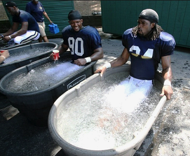
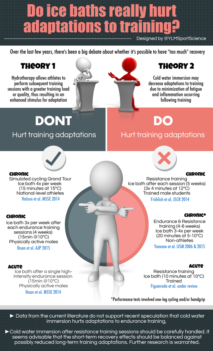
These include nutritional supplements, acupuncture or acupressure, massage, relaxation techniques and hydrotherapy, among others.


Surgery


Joint surgery can repair or replace severely damaged joints, especially hips or knees performed by an orthopaedic surgeon.Surgery can be recommended if pain is very severe or you have mobility problems or if conservative treatments don't help. Some surgical procedure may include osteotomy and arthroplasty.


Helpful site:


Common aches and pain

See more at: http://www.arthritisresearchuk.org/arthritis-information/common-pain.aspx


Exercises to manage pain

See more at: http://www.arthritisresearchuk.org/arthritis-information/exercises-to-manage-pain.aspx